A recently published study in the 'International Journal of Innovative Research in Medical Science' has attempted to formally document reports of anomalous white fibrous structures observed within the circulatory systems of deceased individuals. According to the article, the research corroborates years of anecdotal accounts from embalmers who have encountered obstructions they describe as inconsistent with normal postmortem blood clots.
The survey-based study, led by retired U.S. Air Force Major and data analyst Thomas F. Haviland, alongside Laura Kasner and Daniel Santiago, PharmD, gathered responses over four consecutive years from 2022 to 2025. Participants included embalmers in the United States, Canada, United Kingdom, Australia, and New Zealand, with a total of 808 respondents across the multi-year period.
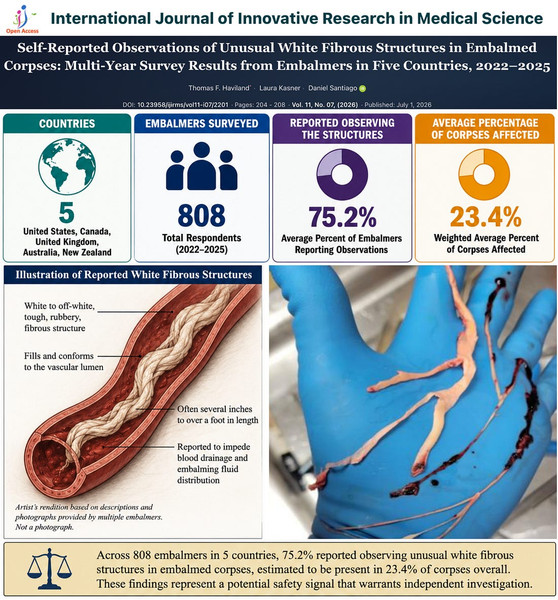
According to the study’s aggregated data, approximately 75.2 percent of participating embalmers reported having observed the white fibrous clots, with the structures estimated to be present in roughly 23.4 percent of embalmed corpses overall. Respondents described the material as typically white or off-white, tough, rubbery, and often measuring several inches to more than a foot in length. Many reported that the structures filled blood vessels, impeded drainage, and interfered with the distribution of embalming fluid. Experienced practitioners characterized them as visually and texturally distinct from the classic “chicken-fat” and “currant-jelly” clots commonly found during embalming procedures.
The study also examined the timing of initial observations. In the 2022 survey, respondents indicated a notable increase in first-time sightings beginning in 2020 and accelerating in 2021. The authors note that this timeframe coincides with the global rollout of COVID-19 vaccination campaigns.
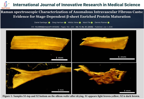
A companion paper published simultaneously moved beyond survey data to laboratory analysis. Led by Santiago, with collaborators including Greg Harrison, Miklós Veres, Mark File, and Dennis Planner, the research employed Raman micro-spectroscopy, protein quantification, and amino-acid profiling to examine representative samples.
The laboratory analysis reportedly identified strong protein signatures, with researchers concluding that the structures represent atypical protein aggregates distinct from conventional postmortem thrombi. Spectroscopic data suggested a structural progression from a native-like α-helical state toward a more advanced β-sheet-enriched configuration, which the authors describe as consistent with stage-dependent protein aggregation. This progressive maturation, they suggest, could account for the structures’ reported length, rubbery consistency, and resilience. While the presence of β-sheet enrichment is commonly associated with amyloid formation, the authors underscored that further testing is necessary before classifying the material as true amyloid fibrils.
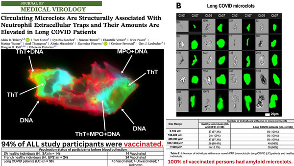
The findings are contextualized within a broader body of research, including a separate peer-reviewed study that reported detecting amyloid microclots in vaccinated individuals. That prior work used Thioflavin-T staining and indicated that purified spike protein could directly induce the formation of insoluble, fibrinolysis-resistant aggregates.
In summation, microscopic amyloidogenic aggregates identified in earlier blood-based research may represent an earlier stage of the same pathological process that produces the large, rubbery fibrous casts reported by embalmers. While the survey and laboratory data are presented as preliminary, the researchers argue that the consistency of reports across multiple countries and the distinct protein signatures identified warrant continued scientific scrutiny rather than dismissal.